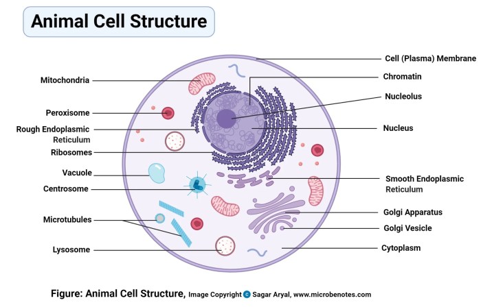
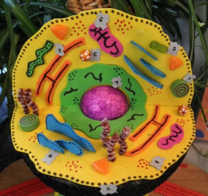
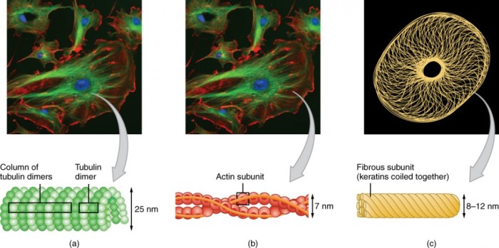
3D Model of Cell, it’s not just a fancy term for a science project. It’s a revolutionary tool that’s changing the way we understand life at its most fundamental level. Imagine peering into the intricate machinery of a single cell, seeing how its parts interact, how it replicates, how it fights off invaders.
This is the power of 3D cell modeling – it brings the invisible to life, offering a level of detail that traditional methods simply can’t match.
These models aren’t just for scientists, either. They’re becoming increasingly important in fields like medicine, where they’re used to design new drugs and therapies, or even in art and design, where they’re inspiring new forms of creative expression. So, buckle up, because we’re about to take a deep dive into the fascinating world of 3D cell models.
3D Models of Cells: A Window into the Microscopic World
The intricate world of cells, the fundamental building blocks of life, has long fascinated scientists and researchers. Understanding the complex structures and processes within these tiny units is crucial for advancing our knowledge of biology, medicine, and even technology.
3D models of cells have emerged as powerful tools, revolutionizing how we visualize, analyze, and interact with the cellular realm.
Importance and Benefits of 3D Cell Models

3D cell models offer a unique advantage over traditional 2D representations. They provide a more realistic and immersive experience, allowing researchers and students to explore the three-dimensional architecture of cells and their intricate components. These models help us understand:
- Spatial organization:3D models reveal the precise arrangement of organelles, proteins, and other cellular structures, providing insights into their interactions and functions.
- Cellular processes:Visualizing processes like cell division, protein synthesis, and signal transduction in 3D helps us grasp their complexity and dynamics.
- Drug discovery:3D models can simulate the effects of drugs on cells, facilitating the development of targeted therapies and reducing the need for extensive animal testing.
- Education and outreach:Interactive 3D cell models provide engaging and accessible learning experiences, making complex biological concepts more understandable for students and the general public.
Types of 3D Cell Models
There are several types of 3D cell models, each with its own strengths and applications:
- Physical models:These are tangible representations of cells, often created using 3D printing or other physical modeling techniques. They are valuable for visualization, teaching, and prototyping.
- Virtual models:These are digital representations of cells, created using computer software. They are highly flexible, allowing for interactive exploration, simulation, and analysis.
- Hybrid models:These combine elements of physical and virtual models, offering the best of both worlds. For example, a physical model could be used for visualization, while a virtual model could be used for simulation.
Creating 3D Cell Models, 3d model of cell
The creation of 3D cell models involves a range of techniques, each with its own advantages and disadvantages:
Microscopy-Based Methods
Microscopy plays a vital role in capturing the intricate details of cells. Several microscopy techniques are used to create 3D cell models:
- Confocal microscopy:This technique uses lasers to scan a sample, producing a series of optical sections that can be stacked to create a 3D image. Confocal microscopy is widely used for visualizing the distribution of fluorescently labeled proteins and organelles.
- Electron microscopy:This technique uses electrons to image a sample, providing extremely high resolution and revealing the fine structure of cells. Electron microscopy is particularly useful for studying the ultrastructure of organelles and macromolecules.
- Light sheet microscopy:This technique uses a thin sheet of light to illuminate a sample, minimizing photodamage and allowing for fast imaging of large volumes. Light sheet microscopy is ideal for capturing dynamic processes in living cells.
Computer-Aided Design (CAD) Methods
CAD software allows researchers to create 3D models from various data sources, including microscopy images, experimental data, and theoretical models. Popular CAD software for cell modeling includes:
- Blender:A free and open-source software that offers a wide range of tools for 3D modeling, animation, and rendering.
- Maya:A professional-grade software widely used in the film and animation industries. It provides advanced features for creating realistic and complex 3D models.
- 3ds Max:Another professional software that is particularly popular for creating architectural and product designs. It also offers powerful tools for cell modeling.
3D Printing Methods
3D printing has revolutionized the creation of physical models, allowing for the fabrication of complex and detailed structures. Several 3D printing techniques are used to create cell models:
- Fused deposition modeling (FDM):This technique involves extruding a thermoplastic filament layer by layer to create a 3D object. FDM is relatively inexpensive and versatile, but the resulting models may have a lower resolution than other methods.
- Stereolithography (SLA):This technique uses a UV laser to solidify a liquid photopolymer layer by layer. SLA produces models with high resolution and smooth surfaces, making them suitable for detailed anatomical models.
- Selective laser sintering (SLS):This technique uses a laser to fuse powdered material layer by layer. SLS is capable of creating complex and intricate models with high strength and durability.
Applications of 3D Cell Models

3D cell models have numerous applications in research, education, medicine, and other fields:
Research and Education
- Drug discovery:3D cell models can be used to screen potential drug candidates, predict their efficacy, and identify potential side effects. This can help accelerate the drug development process and reduce the need for animal testing.
- Cellular mechanisms:3D models allow researchers to study the complex interactions between cells, tissues, and organs. They can be used to investigate the mechanisms of disease, the effects of environmental factors, and the response of cells to various stimuli.
- Diagnostic tools:3D models can be used to develop new diagnostic tools, such as microfluidic devices for detecting biomarkers in biological samples. These tools can improve the accuracy and speed of disease diagnosis.
- Education:3D cell models provide engaging and interactive learning experiences for students of all ages. They can help students visualize complex biological concepts, understand the structure and function of cells, and develop a deeper appreciation for the wonders of the microscopic world.
Medical Devices and Therapies
- Medical device design:3D cell models can be used to design and test new medical devices, such as implants, prosthetics, and drug delivery systems. This can help ensure that these devices are safe and effective.
- Personalized medicine:3D cell models can be used to create personalized medicine approaches, tailoring treatment plans to the specific needs of individual patients. This can improve treatment outcomes and reduce the risk of side effects.
- Tissue engineering:3D cell models can be used to create tissue engineering scaffolds, which provide a framework for cells to grow and form new tissues. This technology has the potential to revolutionize the treatment of a wide range of diseases and injuries.
Other Applications
- Visualization and communication:3D cell models can be used to visualize and communicate complex scientific data, making it more accessible to a wider audience. This can help to improve public understanding of science and promote scientific literacy.
- Art and design:3D cell models have inspired artists and designers, leading to the creation of unique and innovative works of art. These models can be used to explore the beauty and complexity of the natural world.
- Gaming and entertainment:3D cell models are increasingly being used in video games and other forms of entertainment. This can provide players with a more immersive and realistic experience, as well as educate them about the wonders of the microscopic world.
Future Directions

The field of 3D cell modeling is rapidly evolving, with exciting advancements on the horizon. Emerging technologies, such as artificial intelligence and machine learning, are poised to revolutionize the way we create and use 3D cell models. These technologies can help us:
- Automate model creation:AI and machine learning can be used to automate the process of creating 3D cell models from microscopy images and other data sources.
- Improve model accuracy:AI algorithms can be trained to identify and correct errors in 3D cell models, leading to more accurate and realistic representations.
- Develop predictive models:AI can be used to develop predictive models that can simulate the behavior of cells under different conditions, providing insights into disease progression and treatment outcomes.
Despite the exciting advancements, challenges remain in the development of 3D cell models. These include:
- Data availability:The creation of accurate and realistic 3D cell models requires large amounts of high-quality data, which can be challenging to obtain.
- Computational resources:Creating and analyzing complex 3D cell models can be computationally intensive, requiring significant computing power and expertise.
- Standardization:The lack of standardization in 3D cell modeling can make it difficult to compare results across different studies and labs.
Examples of 3D Cell Models
| Cell Type | Modeling Technique | Application | Description |
|---|---|---|---|
| Neuron | Confocal microscopy, CAD | Drug discovery, education | A detailed 3D model of a neuron, showcasing its intricate structure, including the cell body, dendrites, and axon. The model highlights the distribution of key proteins and organelles involved in neuronal function. |
| Epithelial cell | Light sheet microscopy, 3D printing | Tissue engineering, medical devices | A 3D model of an epithelial cell, capturing its characteristic shape and arrangement within a tissue. The model is used to study cell-cell interactions and the formation of epithelial layers. |
| Blood cell | Electron microscopy, CAD | Research, education | A 3D model of a red blood cell, revealing its biconcave shape and the absence of a nucleus. The model highlights the importance of red blood cells in oxygen transport throughout the body. |
Conclusive Thoughts: 3d Model Of Cell
From the tiniest details of cellular structure to the complex processes that govern life, 3D cell models are transforming our understanding of the biological world. With the rapid advancements in technology, we can expect even more incredible breakthroughs in the years to come.
The future of 3D cell modeling is brimming with possibilities, and we’re only just beginning to scratch the surface of its potential.
FAQs
What are the limitations of 3D cell models?
While incredibly powerful, 3D cell models aren’t perfect. They can’t fully replicate the complex environment of a living cell and may not capture all the nuances of cellular behavior.
How are 3D cell models used in drug discovery?
3D cell models allow researchers to test the effectiveness of new drugs and therapies on specific cell types, helping to identify promising candidates and predict potential side effects.
Can anyone create a 3D cell model?
While creating a complex 3D cell model requires specialized skills and equipment, there are also accessible tools and resources available for those interested in exploring this field.